LOCATION AND PERICARDIAL MEMBRANES
The heart is located in the thoracic cavity between the lungs. This area is called the mediastinum. The base of the cone-shaped heart is uppermost, behind the sternum, and the great vessels enter or leave here. The apex (tip) of the heart points downward and is just above the diaphragm to the left of the midline. This is why we may think of the heart as being on the left side, because the strongest beat can be heard or felt here.
The heart is enclosed in the pericardial membranes, of which there are three layers. The outermost is the fibrous pericardium, a loose-fitting sac of strong fibrous connective tissue that extends inferiorly over the diaphragm and superiorly over the bases of the large vessels that enter and leave the heart.
CHAMBERS—VESSELS AND VALVES
The walls of the four chambers of the heart are made of cardiac muscle called the myocardium. The chambers are lined with endocardium, simple squamous epithelium that also covers the valves of the heart and continues into the vessels as their lining (endothelium). The important physical characteristic of the endocardium is not its thinness, but rather its smoothness. This very smooth tissue prevents abnormal blood clotting because clotting would be initiated by contact of blood with a rough surface.
RIGHT VENTRICLE
When the right ventricle contracts, the tricuspid valve closes and the blood is pumped to the lungs through the pulmonary artery (or trunk). At the junction of this large artery and the right ventricle is the pulmonary semilunar valve (or more simply, pulmonary valve). Its three flaps are forced open when the right ventricle contracts and pumps blood into the pulmonary artery. When the right ventricle relaxes, blood tends to come back, but this fills the valve flaps and closes the pulmonary valve to prevent backflow of blood into the right ventricle.
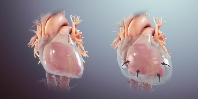
LEFT VENTRICLE
The walls of the left ventricle are thicker than those of the right ventricle, which enables the left ventricle to contract more forcefully. The left ventricle pumps blood to the body through the aorta, the largest artery of the body. At the junction of the aorta and the left ventricle is the aortic semilunar valve (or aortic valve). This valve is opened by the force of contraction of the left ventricle, which also closes the mitral valve. The aortic valve closes when the left ventricle relaxes, to prevent the backflow of blood from the aorta to the left ventricle. When the mitral (left AV) valve closes, it prevents backflow of blood to the left atrium; the flaps of the mitral valve are also anchored by chordae tendineae and papillary muscles.
CARDIAC CYCLE AND HEART SOUNDS
The cardiac cycle is the sequence of events in one heartbeat. In its simplest form, the cardiac cycle is the simultaneous contraction of the two atria, followed a fraction of a second later by the simultaneous contraction of the two ventricles. Systole is another term for contraction. The term for relaxation is diastole. You are probably familiar with these terms as they apply to blood pressure readings. If we apply them to the cardiac cycle, we can say that atrial systole is followed by ventricular systole. There is, however, a significant difference between the movement of blood from the atria to the ventricles and the movement of blood from the ventricles to the arteries.
CARDIAC CONDUCTION PATHWAY
Cardiac muscle cells have the ability to contract spontaneously; that is, nerve impulses are not required to cause contraction. The heart generates its own beat, and the electrical impulses follow a very specific route throughout the myocardium. You may find it helpful you read the following. The natural pacemaker of the heart is the sinoatrial (SA) node, a specialized group of cardiac muscle cells located in the wall of the right atrium just below the opening of the superior vena cava. The SA node is considered specialized because it has the most rapid rate of contraction, that is, it depolarizes more rapidly than any other part of the myocardium (60 to 80 times per minute).
HEART RATE
A healthy adult has a resting heart rate (pulse) of 60 to 80 beats per minute, which is the rate of depolarization of the SA node. (The SA node actually has a slightly faster rate, closer to 100 beats per minute, but is slowed by parasympathetic nerve impulses to what we consider a normal resting rate.) A rate less than 60 (except for athletes) is called bradycardia; a prolonged or consistent rate greater than 100 beats per minute is called tachycardia.
SUMMARY
As you can see, the nervous system regulates the functioning of the heart based on what the heart is supposed to do. The pumping of the heart maintains normal blood pressure and proper oxygenation of tissues, and the nervous system ensures that the heart will be able to meet these demands in different situations. Blood pressure and the blood vessels.